Scientists find that loops of DNA are key to tightly packing genetic material for cell division.
Scientists first discovered chromosomes in the late 1800s, after the light microscope was invented. Using these microscopes, biologist Walter Flemming observed many tightly wound, elongated structures in cell nuclei. Later, it was found that chromosomes are made from DNA, the cell’s genetic material.
Since then, scientists have proposed many possible ways how DNA molecules might fold into 3-D condensed chromosomes. Now, researchers at MIT and the University of Massachusetts Medical School have obtained novel data on the 3-D organization of condensed human chromosomes and built the first comprehensive model of such chromosomes.
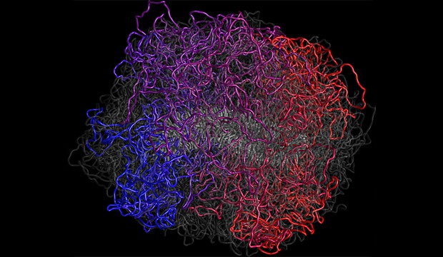
In this model, DNA forms loops that emanate from a flexible scaffold; the loops are tightly compressed along the scaffold. “This is a very efficient way of packing DNA material,” says Leonid Mirny, a senior author of the paper describing the findings.
This condensed state, seen only when cells are dividing, allows cells to neatly separate and distribute their chromosomes so that each daughter cell receives the full complement of genetic material. At all other times, the chromosomes are more loosely organized inside the cell nucleus.
Job Dekker is also a senior author of the paper. Lead authors are Maxim Imakaev, Geoffrey Fudenberg and Natalia Naumova. Other authors are Ye Zhan and Bryan Lajoie.
Layers of structure
Chromosomes are complex molecules with several levels of organization, allowing cells to cram 2 meters of DNA into a nucleus that is only one hundredth of a millimeter in diameter. Long strands of DNA wind around proteins called histones, giving rise to a “beads on a string” structure. Several models have been proposed to explain how those strands of millions of beads are arranged inside tightly packed chromosomes.
“There is no shortage of models of how DNA is folded inside a chromosome,” says Mirny. “Every high-school biology textbook has a drawing of chromosomes folding. If you look at these drawings you might get the impression that the problem has been solved, but if you look carefully you see that all these drawings all very different.”
To help determine which model is correct, the researchers used a technology called Hi-C, which performs genomewide analysis of the proximity of genomic regions. This reveals the frequency of interaction for every pair of regions in the entire genome.
The challenge, however, lies in generating an overall chromosome structure based on Hi-C data. “Given a three-dimensional structure, it is straightforward to find all contacts; however, reconstructing three-dimensional structures from contact frequencies is much more difficult,” Imakaev says.
In 2009, researchers including Imakaev, Mirny, and Dekker used Hi-C to demonstrate that during most of a cell’s life, when it is not dividing, DNA is organized as a fractal globule, in which DNA is not tangled or knotted.
Hi-C also showed that regions with more active genes tend to cluster together in easily accessible compartments, and unused regions form more densely packed clusters. The organization of each chromosome varies among cell types, because every type of cell uses different sets of genes to carry out its function. This means that each chromosome acquires a specific 3-D organization depending on which genes a cell is using.
Chromosomes during cell division
In the new paper, the researchers found that as cells begin to divide, chromosomes are completely reorganized. First, all chromosome-specific and cell type-specific patterns of organization, which are necessary for gene regulation, disappear. Instead, all chromosomes are folded in a similar way as cells begin to undergo cell division, or mitosis. However, the chromosomes do not form the exact same structure every time they condense.
“Unlike proteins, which fold into very defined structures, the chromosomes form a completely different condensed object every time,” Fudenberg says. “It appears similar macroscopically but the individual regions of the genome can be folded in very different ways in different cells.”
The Hi-C technique “provides a modern day molecular microscope, with the power to see inside of these bodies and elucidate their principles of organization,” wrote Nancy Kleckner in a perspective article accompanying the paper. The researchers “combine chromosome conformation capture with polymer physics simulations to provide a new, yet satisfyingly familiar, view,” she wrote.
The researchers believe that two stages are required to achieve the loop-on-a-scaffold structure: First, the chromatin forms loops — each of which contains about 80,000 to 120,000 DNA base pairs — radiating out from a scaffold made of DNA and some proteins. Then, the chromosome compresses itself along its central axis, where the scaffold is located.
While molecular details of the second stage remain mysterious, scientists have a good guess for what might be responsible for the first stage of chromosome folding: A team at Northwestern University recently proposed that proteins called condensins drive chromosome condensation by latching on to the DNA and extruding loops. To test this hypothesis in greater detail, the MIT team is now collaborating with these researchers.
Beyond characterizing condensed chromosomes, this study also opens the door for future work to understand mechanisms of chromosome condensation, cell memory, and epigenetic cell reprogramming.
Content: MIT press release (modified).
Cover image: A model of human mitotic chromosome. The chromosome is composed of a flexible scaffold and a system of loops (red, magenta, blue) - Ilimakaev M., Fudenberg G., Naumova N., Dekker J. and Mirny L.
References
DOI: 10.1126/science.1236083.
Stats
- Recommendations +2 100% positive of 2 vote(s)
- Views 1655
- Comments 0
Recommended
-

Alen Piljić
Managing director | Life Science Network gGmbH
Also:
- President | Research Elements Association
-
amdres rivera
Main position not set