Authors
Alen Piljic
Summary
After spotting plasmid DNA in dishes, cells can be seeded for reverse transfection and semi-automated imaging. This is a continuation protocol to Manual preparation of solutions for spotting DNA by contact printer (for reverse transfection).
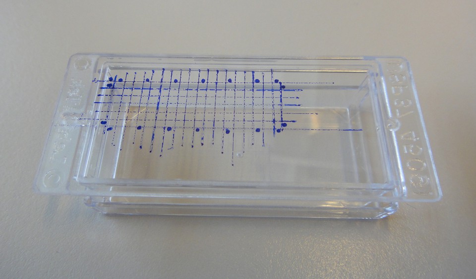 Image 1
 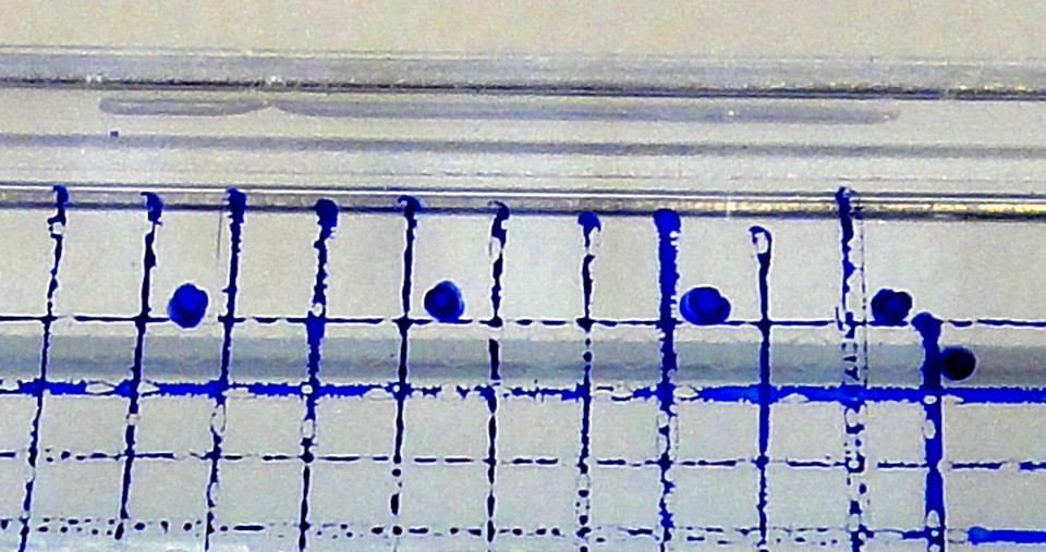 Image 2
 Materials
- cells (e.g. HEK)
- LabTek dishes
- cell culture medium and cell culture equipment
Procedure
- split cells the day before
- mark position of the spots in the dish with a marker (to be able to find them on a microscope)
- seed 100.000 cells (pipet 100uL if in standard splitting there are about 1.000.000 cells/mL) per LabTek dish with 2mL of medium
- drop cells in the middle of the dish, gently distribute
- place dishes in the incubator (the less shaking/vibrations in the incubator the better)
Note: 100.000 cells for seeding is general recommendation; when microscopy is done after 24h, 400.000 – 500.000 cells per LabTek dish may be better
Note2: If cells that like to detach are used, such as HEK, and culture medium needs to be replaced with imaging medium, seed cells in complete medium around noon. Change medium to imaging medium (e.g. optimem without phenol red) after 22h. Perform imaging after at least 2h more incubation at 37C.
Stats
- Recommendations +1 100% positive of 1 vote(s)
- Views 1298
- Comments 0
Recommended
-

Alen Piljić
Managing director | Life Science Network gGmbH
Also:
- President | Research Elements Association