How retroviruses like HIV spread in their hosts had been unknown — until a Yale team devised a way to watch it actually happen in a living organism. The elaborate and sometimes surprising steps the virus takes to reach and spread in the lymph nodes of a mouse have been captured on videos and described in the Oct. 2 issue of the journal Science.
“It’s all very different than what people thought,” said Walther Mothes, associate professor of microbial pathogenesis and co-senior author the paper.
Tracking fluorescently stained viruses in mice, the Yale team led by Mothes and co-senior author Priti Kumar, assistant professor of medicine and microbial pathogenesis, used sophisticated imaging technology to capture the action as the viral particles bind to macrophages via a sticky protein that is located at the capsule of the lymph node.
But that is only the first step of the journey. The captured viral particles open to a rare type of B-cell, seen in red in the accompanying movie. The virus particles then attach themselves to the tail of these B-cells and are dragged into the interior of the lymph node. In one to two days, these B-cells establish stable connections with tissue, enabling full transmission of the virus.
The insights provided by the videos identify a potential way to prevent HIV from infecting surrounding tissue. If researchers could develop a way to block the action of the sticky protein the virus uses to bind to macrophages, then the virus’ transmission could be halted, Mothes suggested.
“The direct study of viral pathogenesis within living animals should reveal more surprises in the future,” Mothes said.
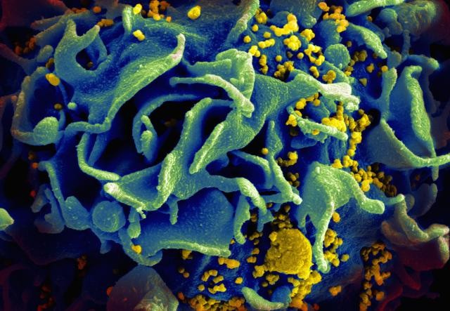
Cover image: Scanning electromicrograph of an HIV-infected T cell (NIAID, Flickr).
References
DOI: 10.1126/science.aab2749.
Stats
- Recommendations +1 100% positive of 1 vote(s)
- Views 8447
- Comments 0
Recommended
-

Alen Piljić
Managing director | Life Science Network gGmbH
Also:
- President | Research Elements Association